
Celebrating the next generation
“That was probably the last exam of your life,” said Professor Michael Gotthardt in his speech at the PhD graduation ceremony in November. Some 50 doctoral students successfully completed their PhD at the Max Delbrück Center for Molecular Medicine in the Helmholtz Association (MDC) in 2019. “It is a good reason to celebrate,” noted Gotthardt, who served as the master of ceremonies for the event. “You have now closed an important chapter in your lives.” The graduation ceremony for the young researchers took place for the first time in the MDC’s new building in Berlin-Mitte, home to the Berlin Institute for Medical Systems Biology (BIMSB).
You have now closed an important chapter in your lives.
Many of the freshly minted PhDs have already left Berlin behind to work at research centers around the world. Dr. Verena Haage, who conducted research on microglia cells in Professor Helmut Kettenmann’s lab, is also relocating to a distant place. She reported after the ceremony that she will be working at Columbia University next year. Others, such as Dr. Petar Glazar, will remain at the MDC for a while longer: “I’m going stay another year in Nikolaus Rajewsky’s lab in order to pursue questions that arose from the findings of my dissertation,” said Glazar, who completed his PhD on circular RNA.
In 2019, about 50 doctoral students completed their doctorates at the Max Delbrück Center for Molecular Medicine in the Helmholtz Association (MDC). For the first time, the closing ceremony took place at the BIMSB.


Four PhD prize winners

PhD Publication Prize winner Karlien Yvonne Debus presented her research at the PhD graduation ceremony 2019.
The annual PhD Prize, sponsored by the Society of Friends of the MDC, was presented in recognition of the best publications. The first prize of €1,000 went to Karlien Yvonne Debus, a doctoral student from Professor Gary Lewin’s lab. Debus presented the findings of her work at the ceremony. Her research focuses on why highveld mole rats, an African rodent, don’t feel pain when stung by some venomous insects. She found out that a particular gene is very active in the rodents. This gene, she explained, is responsible for pain insensitivity to certain substances. Over the course of evolution, this has given the mole rats an advantage over other rodents: They share their natural habitat, underground burrows, with venomous ants whose stings don’t bother them. Debut, one of two lead authors of the paper, published the findings in 2019 in the journal Science.
The papers by doctoral students Jonathan Alles, Eleni Kabrani, and Daniel Wenz were honored with second place, with each young researcher receiving a prize of €250. Alles was involved in the development of FLAM-seq in Nikolaus Rajewsky’s lab. This method enables full-length mRNA sequencing including their poly(A) tails. This was only possible previously by using complicated algorithms and only on a limited basis. Messenger RNAs (mRNAs) translate the instructions present in DNA into proteins. The length of their tails plays an important role in the regulation of genes. Alles was one of the lead authors who published the method in the journal Nature Methods in 2019.
Eleni Kabrani, another second-place winner, investigated in Professor Klaus Rajewsky’s lab the role played by a certain transcription factor in Burkitt’s lymphoma, a cancer that develops from a particular type of immune cell. Many of these tumor cells have mutations that cause the transcription factor to be missing from its normal place in the cell. It usually regulates signaling pathways that destroy malignant cells or repair DNA. Kabrani showed that in this type of cancer the transcription factor has the exact opposite effect: It helps the tumor grow and survive. The findings were published in the journal Blood.
The paper by Daniel Wenz, which appeared in the journal Magnetic Resonance in Medicine, was also recognized. For the first time, researchers imaged the distribution of potassium in a human heart without medical intervention. This was made possible by ultrahigh field magnetic resonance (UHF MR) imaging. MR technologies use a strong magnetic field and radio waves to generate slice-by-slice images of organs. Important components of MR scanners are antennas and detectors that send and receive radio waves. This is comparable to the functionality of a radio or mobile phone. Daniel Wenz from Professor Thoralf Niendorf’s lab designed an antenna system for the MDC’s 7-Tesla scanner that allows researchers to measure very weak potassium signals to image their spatial distribution in the heart. This approach enables to investigate changes in potassium concentrations and their influence to heart function.
„Best Scientific Image“
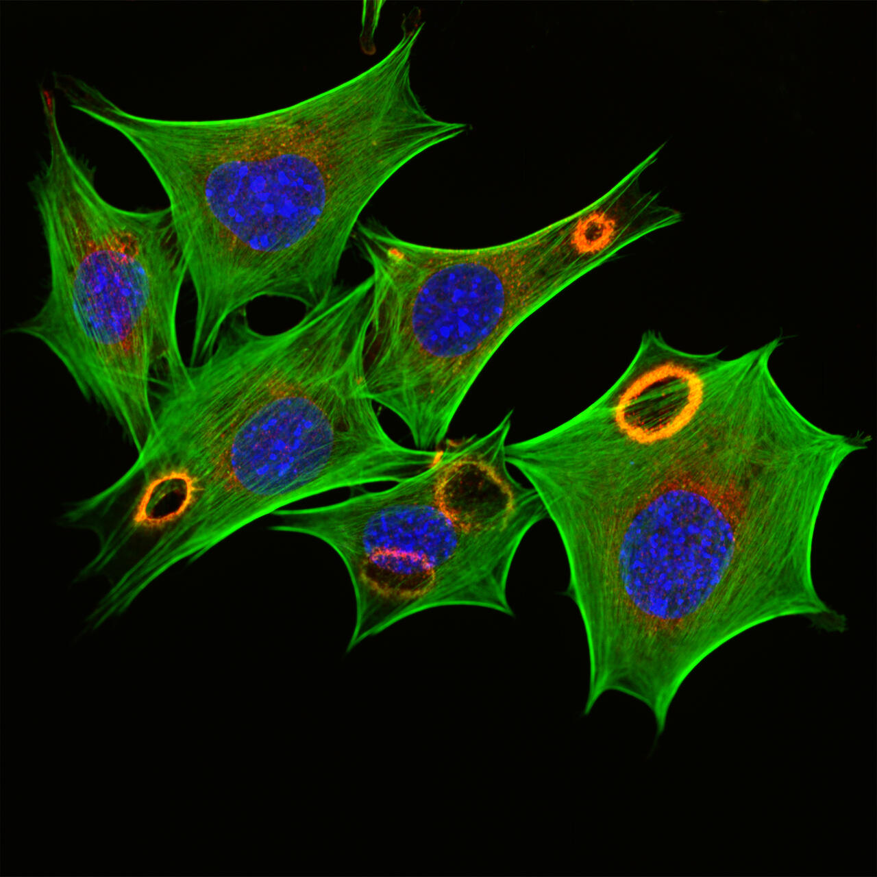
Fibroblasts, specific cells of the connective tissue stimulated with growth factors, can be seen. Growth factors are released in injuries from blood platelets, among other things, to stimulate tissue healing. Fibroblasts are stimulated to divide, migrate into wounds and produce collagen. Fluorescence staining shows the actin cytoskeleton (green), the cell nucleus (blue) and the protein cortactin (red). Cortactin is located at the dorsal (upper) cell membrane in ring-shaped vertical waves that spread like a fire and transform the actin cytoskeleton within a few minutes.
The Best Scientific Image Award has been presented since 2007. This year, the award committee received some 20 entries from employees at the Buch campus. Visitors to the Long Night of the Sciences in June 2019 voted for the best image. Kettenmann announced the winners at the PhD graduation ceremony. First place went to Fabian Lukas from the Leibniz-Forschungsinstitut für Molekulare Pharmakologie. His image shows connective tissue cells which were stimulated with growth factors. Several MDC scientists took second place: Dr. Hanna Napieczynska from Dr. Arnd Heuser’s lab participated with an image that depicts the blood vessels of a mouse kidney. It was captured using high-resolution computed tomography. Charlene Memler, Dr. Florian Rau, Dr. Svenja Steinfelder, and Dr. Clarissa Whitmire, all from Dr. James Poulet’s lab, submitted an image in which the sensory receptors on a mouse’s paw can be seen. A heart-brain-organoid, photographed by Dr. Sebastian Diecke from the Berlin Institute of Health (BIH), was selected as third prize. The Society of Friends of the MDC and Nikon sponsored the prizes for the winners.


