
The fly atlas

Robert Zinzen is head of the lab "Systems Biology of Neural Tissue Differentiation".
Scientists have been using the fruit fly Drosophila melanogaster as a model organism for over 100 years, for example to study how diseases develop. In 2017, Drosophila researchers from all over the world joined forces to produce a comprehensive cell atlas for the species. They recently published an atlas of more than 580,000 cells from all parts of the animal's body in Science. The atlas serves as a resource for the entire Drosophila research community, supporting work such as the study of genetic disorders and cellular processes in a complete organism with single-cell resolution. In this interview, Dr Robert Zinzen from the Max Delbrück Center for Molecular Medicine in the Helmholtz Association (MDC) explains how this unusual mapping project came about and what its purpose is. Zinzen, one of the initiators of the project, leads the Systems Biology of Neural Tissue Differentiation lab at the Berlin Institute for Medical Systems Biology (BIMSB), part of the MDC.
When I hear the word 'atlas', I think of a book with lots of maps. What does an atlas of fly cells look like?
There are different kinds of atlases. Generally speaking, they provide an overview of particular places or situations and contain lots of different information. The Fly Cell Atlas provides a complete picture of all the cells found in Drosophila. This is because to understand how an organism works, we need in-depth knowledge of its cellular components.
Does the atlas show where the various cells are in the organism?
We don't yet have a complete spatial resolution, as you would with a map – it's more like a catalogue of over 250 different types of cell, which shows the tremendous diversity. Although we can compare the heart and the wing, for example, in both we find the same cell types, such as nerve cells; and we don't yet know exactly where the individual cells are located.
What are the advantages of having a list of cell types like this?
We want to find out how the cells tick, and to do that we need to characterise them in more detail. For example, how do muscle cells in different types of tissue differ from each other? What is their genetic signature? What genetic switch activates which gene? What biochemical repertoires do the cells have? There are lots of different molecules that determine the specific characteristics and abilities of a cell: signalling molecules and transcription factors, for example, which control protein production. We also find rare cell types and unusual subtypes.
And the atlas brings all this information together in one place?
Exactly. It gives us a complete picture of all cells in the fly organism. Once we have this information, we can then study how the cells interact and communicate with each other, for example. We can do this without knowing their exact position. But at some point we do want to complete the spatial resolution. That will be the next big step.
How will you achieve that?

Scientists in the Zinzen lab work with flies.
There are various approaches we can use. One is a computer-based method that reconstructs the spatial resolution from the available data. Another method determines the position of the cells directly through sequencing experiments. Both approaches have their advantages and drawbacks. But the important thing is that methods do exist that will eventually make it possible.
Why an atlas of a tiny fly and not a more 'interesting' organism?
As a matter of fact there are other, similar projects going on, including the Human Cell Atlas. But because of the fly's size, it's less complex, so we can tackle it more comprehensively. It's the first time that a complete organism has been mapped in this level of detail. So for the first time we can study how it functions in its entirety.
And this new knowledge can then be transferred to humans?
To a large extent, yes. That's why we call the fly a model organism. There are numerous principles that apply equally or in a similar way in the human body as in the fly. It could be the tasks performed by genes or cells, or disease models. For over 70% of genes associated with human diseases there is a direct counterpart in the fly genome.
Who can use the atlas?
In principle, anyone who is interested. From the outset we worked to ensure that the data was freely available. But not everyone can work with such huge amounts of data. So we also provide processed files in which the information is presented visually. But obviously it's intended primarily for the Drosophila research community, for whom the atlas provides a unique platform for studying and understanding biological mechanisms.
Why is the volume of data so large?
The map of the adult fly that we present in the new paper consists of more than half a million cells. And each one contains information about thousands of genes and transcription factors. It's a huge, multidimensional dataset. To analyse it you need various computer-based methods, including machine learning technologies. These are algorithms that learn autonomously and find patterns in the data, for example to classify cells.
You were one of the initiators of the project. How did that come about?
Fruit flies have played a leading role in biological research for over a century. With their help, Thomas Hunt Morgan discovered that genes sit on chromosomes.
The idea of a fly cell atlas came about in 2015 and 2016 as a result of the development of techniques for sequencing individual cells, something that the MDC played an important role in – especially the lab headed by Professor Nikolaus Rajewsky. Once we could do single-cell sequencing, suddenly generating the data was no longer the problem. At the time my lab was already working on the fly embryo, which consists of around 6,000 cells. Many characteristic gene expression patterns can be seen at a very early stage of development. We collaborated with the Rajewsky lab to try and work out the spatial position of the cells from the experimental data using mathematical methods.
This was the first attempt at mapping?
Yes, if you like. Internally, we called it the embryo's 'blueprint'. Together with Drosophila experts Professor Stein Aerts from the Flemish Institute for Biotechnology in Belgium and Professor Bart Deplancke from EPFL in Lausanne, Switzerland, I decided to set up a consortium to create a complete cell atlas of the fly. In 2017, we organised our first conference, with around 40 to 50 people, in Leuven in Belgium. We discussed the relevant issues and eventually laid the foundations for the project. Last year, we also met here at the MDC in Berlin to discuss how things were going. We've become a huge international community. You can see this in our latest publication, which is the joint effort of more than 150 people from 46 different research groups based in many different countries.
Interview by Janosch Deeg

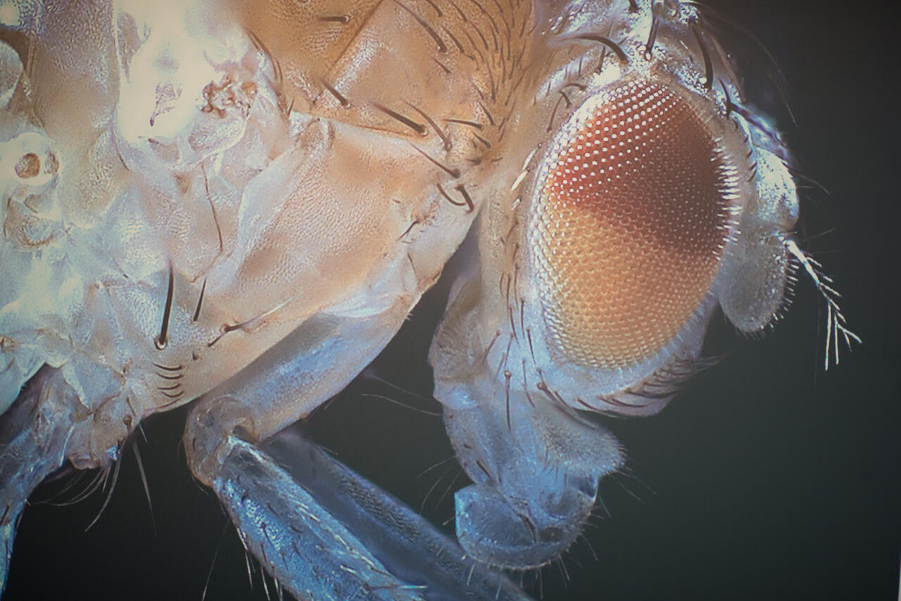
Drosophilidae are a popular research model organism.
Further information
- What flies and humans have in common
- Press release by VIB-KU Leuven Center for Brain and Disease Research
Literature
Hongjie Li (2022): “Fly Cell Atlas: A single-nucleus transcriptomic atlas of the adult fruit fly“. Science. DOI: 10.1126/science.abk2432


